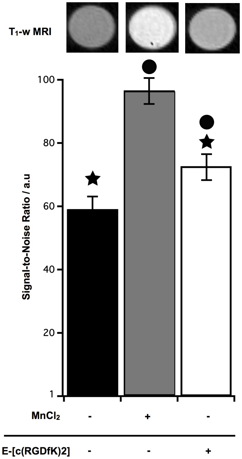
Figure 1. MRI signal of U87 cells after uptake of E-[c(RGDfK)2]-DOTA-Gd.

U87 cells were incubated at 7*104 cell/cm2, with E-[c(RGDfK)2]-DOTA-Gd or MnCL2 (400 µM during 4 h and 1 h, respectively). Applying a T1-weighted sequence (T1-w MRI), results with E-[c(RGDfK)2]-DOTA-Gd were significantly different from the unlabeled cells (★, p<0.05) and from the MnCl2 condition (•, p<0.05).
