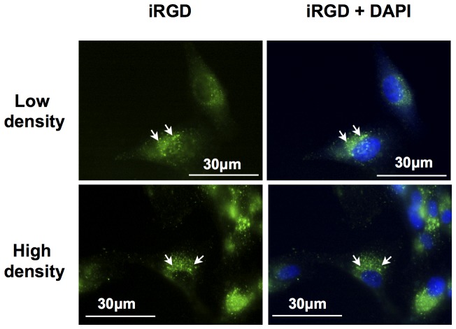
Figure 3. Observation by fluorescent microscopy of the iRGD-alexa488 internalization into U87 cells.
U87 cells were incubated during 1h at 37°C with iRGD-alexa488 at 15 µM (green). Nuclei were stained with DAPI (Blue). Arrows showed vesicular accumulation of the fluorescence inside the cytoplasm. The cytoplasmic membrane labeling was not detectable.

