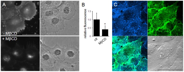
Figure 3. Cell surface labeling with TNM-AMCA.
(A, B) A431 cells were fixed and labeled with TNM-AMCA. The effect of sterol extraction with MβCD was examined. Fluorescent images (A) and the quantification of the fluorescence (B) are shown. Relative fluorescence corresponds to the mean ± SD for more than 35 cells in two independent experiments. Asterisk indicates statistically significant difference (p<1×10⁻¹⁰). (C) HeLa cells were co-stained with TNM-AMCA (blue) and BCθ (green). Merged fluorescence (lower left) and DIC (lower right) images are also shown. Cells were fixed, incubated with BCθ followed by streptavidin-Alexa Fluor 488 treatment, and then labeled with TNM-AMCA. Scale bars, 50 µm.

