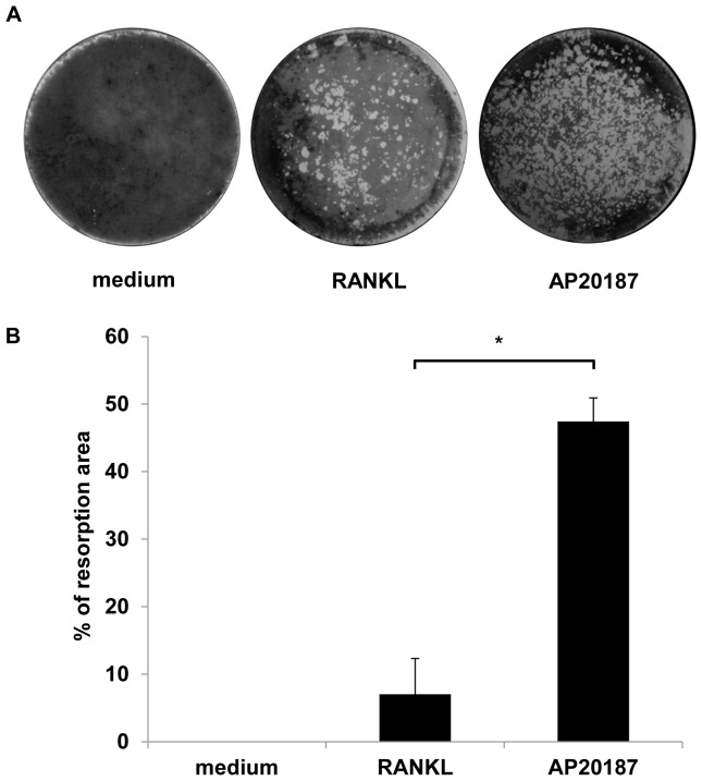
Figure 6. CID induced osteoclasts resorbed a two-dimensional mineralized substrate.
(A) RAW264.7+iRANK cells were treated with either RANKL (100 ng/ml) or AP20187 (100 nM) on Osteologic discs. After 10 days, resorption lacunae were visualized by von Kossa staining. (B) The percent resorbed area per disc was measured and analyzed using ImageJ. Three experiments were averaged. Y-axis shows the % resorption per disc. *p<0.05.

