
Figure 2. Specific and selective uptake of C2IN-C3lim into macrophage-like RAW 264.7 cells.
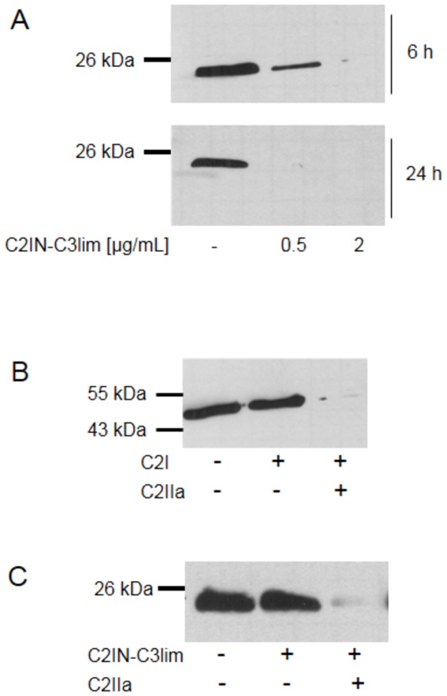
A. ADP-ribosylation status of Rho in RAW 264.7 cells treated with C2IN-C3lim. Cells were incubated with 0.5 or 2 µg/mL of C2IN-C3lim or left untreated for control. The cells were lysed after 6 and 24 h and equal amounts of lysate proteins incubated with fresh C3bot1 and biotin-labelled NAD+. The biotinylated, i.e. ADP-ribosylated Rho is shown. Equal amounts of loaded protein were confirmed by Ponceau S staining of the blotted proteins (not shown). B. C2I alone is not taken up into RAW 264.7 cells. Cells were incubated with C2I (2 µg/mL) alone or with C2I (0.4 µg/mL) + C2IIa (0.8 µg/mL) or left untreated for control. After 6 h cells were lysed and equal amounts of lysate proteins incubated with fresh C2I and biotin-labelled NAD+. The biotinylated, i.e. ADP-ribosylated actin is shown. Equal amounts of loaded protein were confirmed by Ponceau S staining of the blotted proteins (not shown). C. C2IN-C3lim is not taken up into pre-osteoblastic MC3T3 cells under comparable conditions. Cells were incubated with C2IN-C3lim (5 µg/mL) or with C2IN-C3lim (1 µg/mL) + C2IIa (2 µg/mL) or left untreated for control. After 6 h the cells were lysed and the ADP-ribosylation status of Rho determined as described in A. The biotinylated, i.e. ADP-ribosylated Rho is shown. Equal amounts of loaded protein were confirmed by Ponceau S staining of the blotted proteins (not shown).