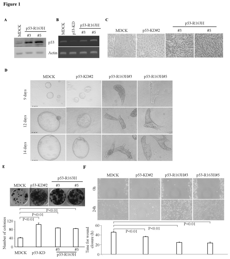
Figure 1. Overexpression of mutant p53 R163H disrupted tubular formation in 3-D culture.
A, Generation of MDCK cell lines in which siRNA-resistant mutant p53-R163H was stably overexpressed (clones 3 and 5). The level of p53-R163H was determined by Western blotting. B, The level of wild-type p53 transcripts was determined by RT-PCR. C, Representative images of MDCK cells, MDCK cells with p53 knockdown, or MDCK cells with mutant p53 (R163H) in 2-D culture (200×). D, Representative images of MDCK cells, MDCK cells with p53 knockdown, or MDCK cells with mutant p53-R163H in 3-D culture for 6 d or 12 d. Scale bar: 100 µM. E, Top panel: colony formation assay was performed with MDCK cells, MDCK cells with p53 knockdown, or MDCK cells with mutant p53-R163H. Bottom panel: the number of colonies was counted and presented as Mean ± SD from three separate experiments. F, Wound healing assay was performed with MDCK cells, MDCK cells with p53-KD, or MDCK cells with mutant p53-R163H. Top panel: cell migration was determined by visual assessment of cells migrating into the wound for 24 h using a phase-contrast microscopy. Bottom panel: the time required for wound closure was measured and presented as mean ± S.D. from three separate experiments.

