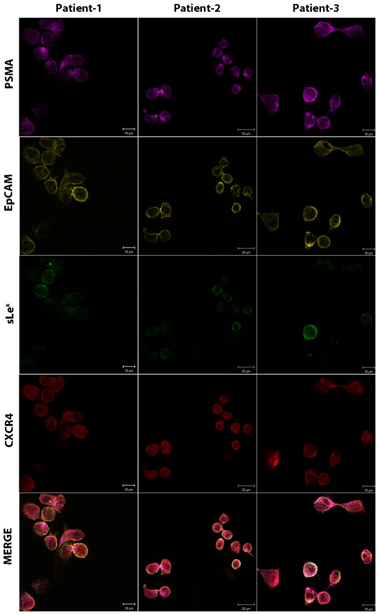
Figure 3. Isolation of prostate CTCs from metastatic PCa patients using anti-CD45 immunomagnetic depletion.

2.5 ml blood from three metastatic PCa patients (> 50 CTCs/ 2.5 ml blood) was processed via ficoll density centrifugation and the PBMC fraction was collected. Immunomagnetic anti-CD45 depletion was performed on the obtained PBMCs and the remaining cells were washed, cytospunned onto the slides. Slides were stained for PSMA, EpCAM, sLex, and CXCR4 using the protocol as described in Figure S1. MDA, PC3, and KG1 cells were simultaneously stained as a control for the following markers: PSMA= Magenta, EpCAM= Yellow, HECA-452= Green, CXCR4= Red. All prostate CTCs expressed CXCR4, while, sLex expression was variable. The analysis of sLex intensity is shown in Figure 4.
