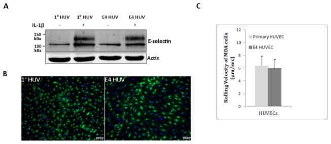
Figure 5. E-selectin expression and its functional assay in primary and E4ORF1 HUVECs.

A) Western blot showing E-selectin protein expression in primary (1°) and E4ORF1 HUVECs. HUVECs were either stimulated with 50 ng/ml IL-1β for 4 h or left untreated. E-selectin= 95-115 kDa, based on post-translational modifications. The blot underneath E-selectin shows actin, used as a loading control. Western blot is a representative of three independent experiments. B) Immunofluorescence showing E-selectin expression in IL-1β-stimulated 1° HUVECs (passage=2) and E4ORF1 HUVECs (passage 10). E-selectin= Green, DAPI= Blue. C) Rolling velocity of 106 MDA cells on 50 ng/ml IL-1β-stimulated 1°- and E4ORF1- HUVECs. HUVECs were stimulated with IL-1β for 4 h. The mean rolling velocity of MDA cells at 1 dyn/cm2 was 5.94 + 3.43 µm/sec and 6.35 + 3.92 µm/sec on E4-ORF1 and primary HUVECs, respectively. No significant difference was seen in the rolling velocities of MDA cells on either 1° or E4ORF1- HUVECs. Graph depicts Mean + SD.
