Figure 1.
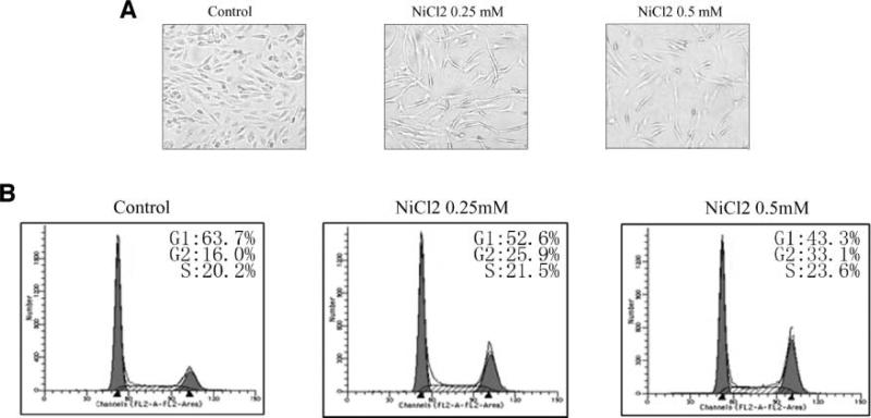
Nickel-triggered G2/M cell cycle arrest and proliferation blockage. 1 × 10³ of Beas-2B cells were seeded into each well of 96-well plates, cultured in 10% FBS DMEM overnight, and then exposed to NiCl2. The cells were photographed under microscopy after 36 h exposure (A). Beas-2B cells were seeded into each well of 6-well plates and cultured in DMEM containing 10% FBS. After the cell density reached 70% to 80%, the cells were exposed to 0.25 mmol/L or 0.5 mmol/L NiCl2 for 36 h, and then were fixed and stained with propidium iodide as described previously. Cell cycle distribution was determined by flow cytometry (B). Each experiment was repeated for at least three times.

