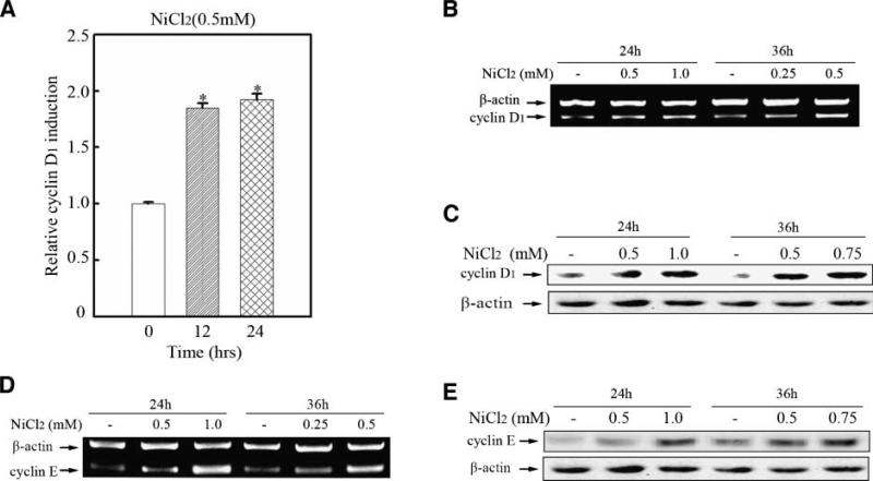
Figure 2.
Nickel up-regulated cyclin D1 and cyclin E expression. 8 × 10³ Beas-2B cyclin D1-luc mass1 cells (A) were seeded into each well of a 96-well plate. After being cultured at 37°C overnight, the cells were treated with 0.5 mmol/L NiCl2 for various time periods as indicated. The cells were then extracted with lysis buffer, and the luciferase activity was measured as described in Materials and Methods. Bar, mean and SD of the triplicate wells; (*), a significant increase from medium control cells (P < 0.05). Beas-2B cells (B, D) were seeded into 100-mm dishes. After being cultured at 37°C overnight, they were treated with NiCl2 for 24 or 36 h as indicated. RNA isolation and reverse transcription-PCR (RT-PCR) were carried out as described in Materials and Methods. 2.5 × 10⁵ Beas-2B cells (C, E) were seeded into each well of 6-well plate and cultured in 10 % FBS DMEM. Twenty-four hours later, the cells were exposed to NiCl2 at various dosages for 24 or 36 h as indicated and then Western blot assay was done.

