
Figure 6.
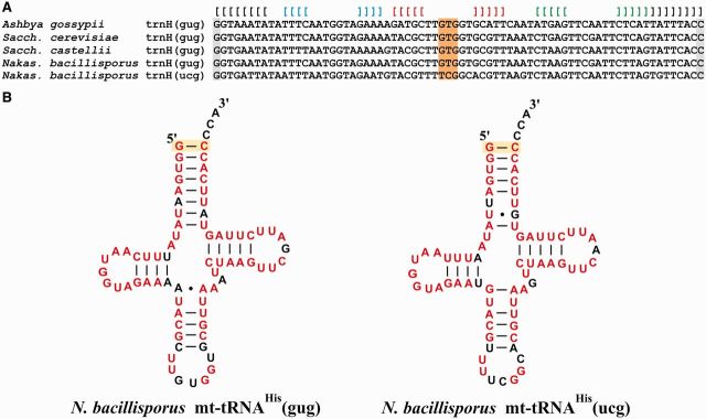
Primary and secondary structures of N. bacillisporus mitochondrial  and putative
  . (A) Selected yeast mitochondrial tRNAsHis with GUG anticodons (anticodon marked orange) are aligned in comparison with an unusual, predicted N. bacillisporus mitochondria tRNAHis with a UCG anticodon (that according to the standard genetic code would recognize CGN arginine). Square brackets indicate the four typical helical regions in tRNAs. Note the characteristic G residue at position −1 (constituting the 5′ terminus of tRNA histidine) that pairs with the C at the 3′ discriminator position (both positions marked in gray). (B) Secondary structures of the two mitochondrial N. bacillisporus histidine tRNAs. Nucleotide identity is coded in red, and the recognition sequence for HisRS (G–C base pair at position −1) is marked yellow.