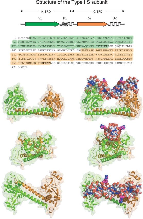
Figure 4.

Structure of the Type I S subunit (pdb:1YF2). The recognition sequence of this protein, S-MjaXI, from Methanocaldococcus (formerly Methanococcus) jannaschii is not known. It is closely related to the EcoKI-family (Type IA) of enzymes depicted in Figure 1. The upper diagram shows the domain organization of the protein; arrows represent DNA-binding domains, and curly lines represent dimerization alpha helices. The aa sequence of the protein is shown below, with the domains in corresponding colors. Below this are three views of the structure, from three perpendicular directions, ‘sideways’, ‘end-on’, and ‘above’. The panels on the left depict the protein; those on the right depict the protein with modeled DNA positioned approximately as it is bound. The DNA was taken from pdb:2Y7H and transferred by structural alignment of the S subunits.
