
Figure 4.
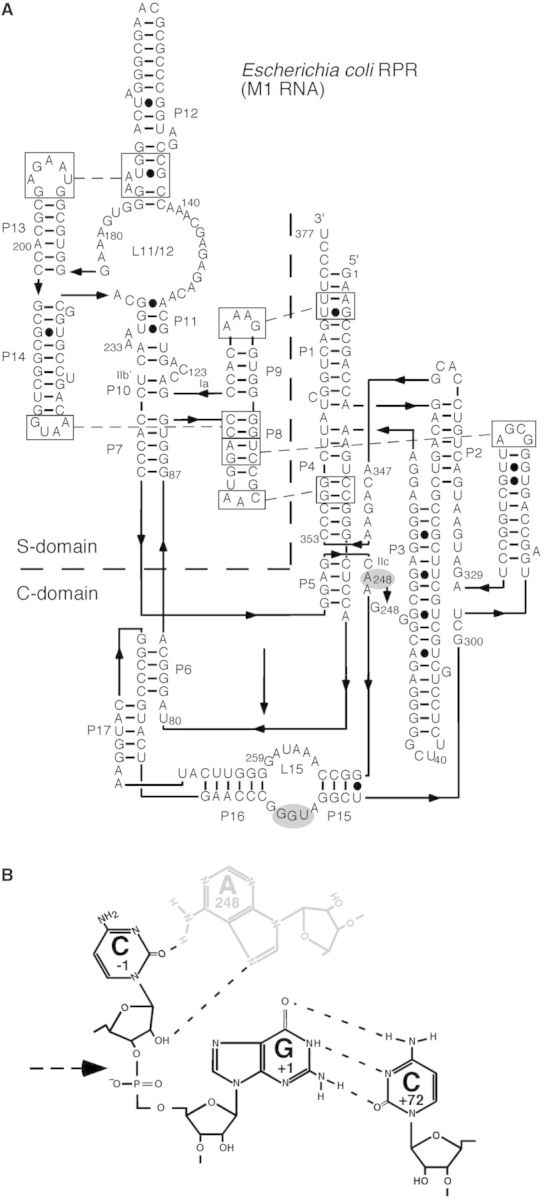
Secondary structure model of Eco RPR and a model of the N−1/A248 interaction. (A) Secondary structure of Eco RPRwt according to Massire et al. (39). Residue A248 that was changed to G is indicated in grey. (B) A putative model of the N−1/A248 interaction. Black and grey residues mark substrate and RPR residues, respectively, and the dashed arrow marks the scissile bond.