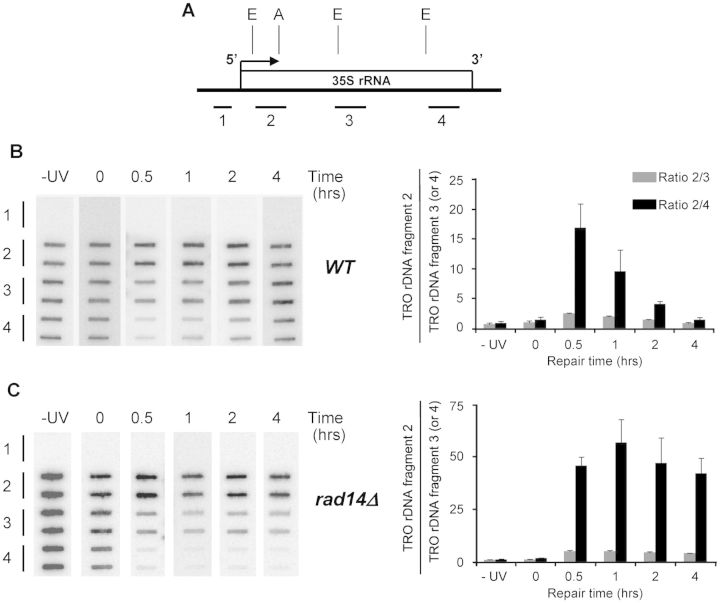
Figure 4.
RNAPI elongation in wild-type (WT RAD14) and rad14Δ cells before and at different times after UV irradiation, at the 5′-end, middle and 3′-end of rRNA gene. (A) EcoRI (E) and ApaI (A) are shown as references to Supplementary Figure S1 and Figures 1A and 5A. Bars position the rDNA fragments: 1; 532 bp, 2 to 4; average length of ∼950 bp. (B) Left panel; nuclei from nonirradiated (−UV) and irradiated (0, 0.5, 1, 2 and 4 h) WT were incubated in TRO reaction conditions containing [α-32P]UTP. Purified elongated, radio-labeled RNAs were used as probes to hybridize the membrane-bound rDNA fragments (1–4), spotted in duplicate onto filter membranes. Fragment 1 was the control for hybridization specificity. Right panel; quantification of phosphoimager signals. Histogram shows measurements for the 5′-end (rDNA-fragment 2) either normalized to the corresponding measurements for the middle (rDNA-fragment 3, gray bars) or the 3′-end (rDNA-fragment 4, black bars). Means (±1 SD) of three independent biological experiments. (C): Same as in (B) for rad14Δ.

