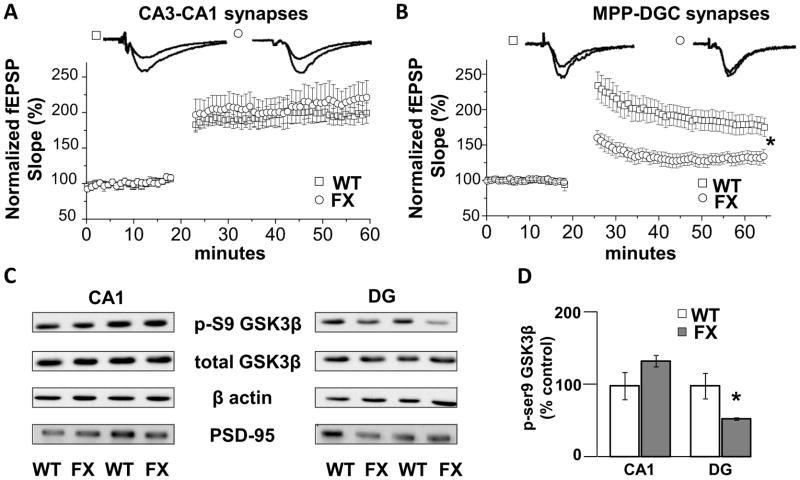
Figure 1.
Deficits in LTP at MPP-DGC synapses in FX mice are accompanied by decreased inhibitory serine-phosphorylation of GSK3β. (A) Summary plots of the magnitude of LTP induced by HFS at CA3-CA1 Schaffer collateral synapses in slices from WT (n=7) and FX (n=6) mice. (B) Summary plots of the magnitude of LTP induced by HFS at MPP-DGC synapses in slices from WT (n=6) and FX (n=9) mice. (C) Representative Western blots showing a reduction in phospho-serine9-GSK3β (p-S9 GSK3) protein in dentate gyrus (DG) but not in CA1 from FX versus WT mice. Total GSK3β protein levels are not different between groups. β-Actin and PSD-95 were used as cytosolic and synaptic protein loading controls. (D) Quantitation of the ratio of p-S9 GSK3β to total GSK3β normalized to values of WT mice (CA1, n=4, and DG, n=6, for each genotype). *p<0.05 (Student’s t test).

