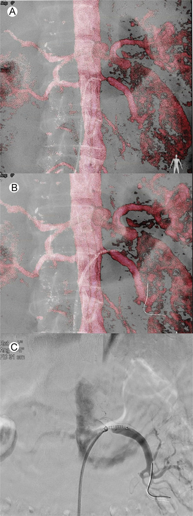
Figure 4.
Renal artery stenting CBCT image fusion workflow. (A) Catheterization of the left renal artery using the volume-rendering MRA overlay. The tip of the catheter is in the ostium of the stenosed renal artery. (B) Placement of the stent under VR MRA overlay to cover the entire lesion. No contrast was used till this part of the procedure. (C) To confirm stent position in relation to stenosis prior to final deployment, 3 cc of contrast was injected. (Color version of figure is available online.)

