
Figure 4.
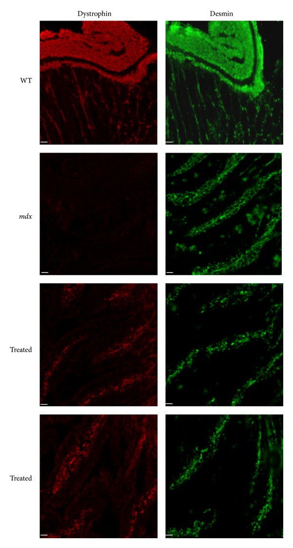
Immunofluorescence analysis of intestinal smooth muscle of wild type (WT), untreated (mdx) and alginate-ZM2-M23D orally treated (Treated) mdx mice. The sections of small intestine were labeled with antidystrophin antibody. Serial sections of intestine were labeled with a polyclonal antibody for desmin (green). All samples were observed with a Nikon Eclipse 80i fluorescence microscope. Dystrophin (red) is clearly visible in the intestinal smooth muscle of WT mice, absent in untreated mdx, and rescued in treated mdx mice. (Scale bar = 50 μm).