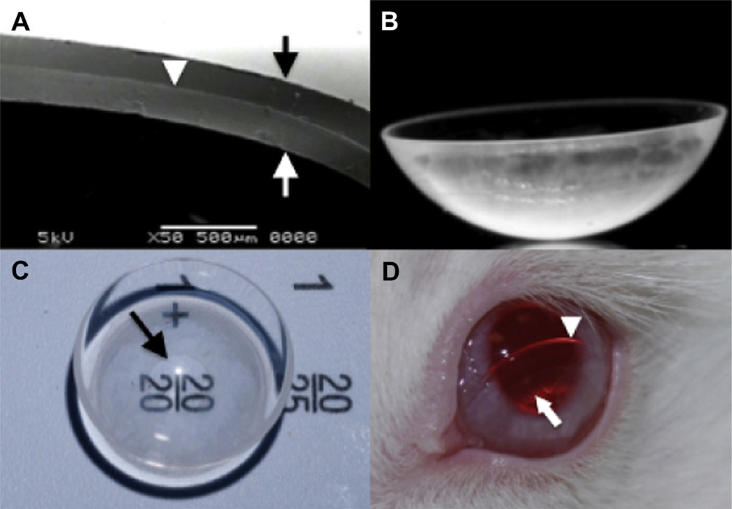
Fig. 1.
The latanoprost-eluting contact lens. (A) Representative scanning electron microscope (SEM) image of a cross-section of the lens demonstrated the drug–polymer film (white arrowhead) located between the methafilcon hydrogel’s outer surface (black arrow) and inner surface (white arrow). (B) Photograph from side view. (C) Top view shows a clear central aperture surrounded by a translucent ring of drug–polymer film. Arrow points to the inner margin of the drug polymer film. (D) The contact lens on the surface of a rabbit’s eye. The lens was intentionally displaced inferiorly for better visualization of the lens edge (white arrowhead) and inner diameter of the drug–polymer film (white arrow).

