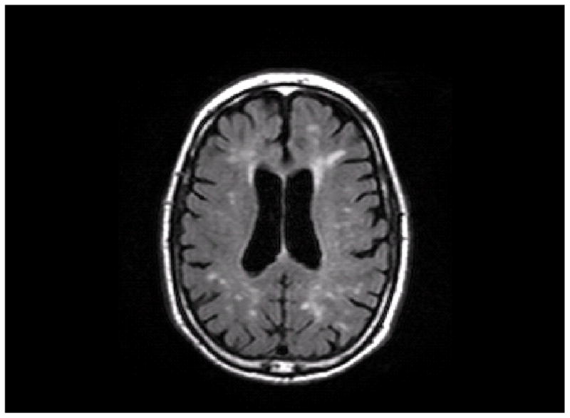
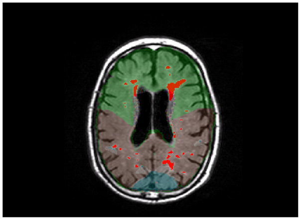
Figure 2.


An axial slice from a T2-weighted FLAIR image. A, This image shows the unlabeled MRI scan. B, This image shows WMH labeled in red and a lobar atlas superimposed on the image. Frontal lobe is labeled in green, parietal lobe is labeled in brown, and occipital lobe is labeled in blue (temporal lobe is not visible at this level).
