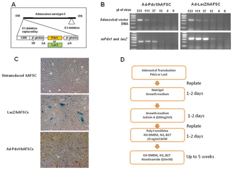
Figure 1.
AFSCs transduction with a Pdx1-expressing adenovirus and the transgene expressed. (A) Schematic representation of the adenoviral vector (35 840 bp) containing the mouse Pdx1 cDNA. CMV, cytomegalovirus promoter. (B) (top) RT–PCR of adenoviral vector DNA at different amounts (ml) used to transduce AFSCs; (bottom) RT–PCR of mPdx1 and lacZ genes at different amounts (ml) used to transduce AFSCs. (C) LacZ and PDX1 protein expression in transduced AFSCs, as determined by hydrolysis of X-Gal substrate (middle) and immunostaining of mPdx1 (right). (D) A scheme showing the sequence of hAFSCs viral transduction and culture conditions during pancreatic differentiation

