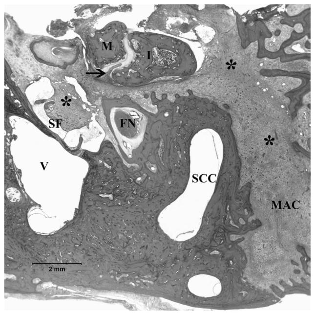
FIG. 1.
In the right temporal bone, the middle ear and mastoid cells were filled with pathologic tissue (asterisk), which is characteristic of Langerhans’ cell histiocytosis. Incudomalleolar joint invasion by Langerhans’ cell histiocytosis (arrow) (hematoxylin and eosin; original magnification, ×1). FN indicates facial nerve; I, incus; M, malleus; MAC, mastoid air cells; SCC, semicircular canal; SF, stapes footplate; V, vestibule.

