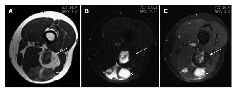
Figure 2.

Myxoid liposarcoma. A: Axial T1-weighted image; B: Fat-saturated T2-weighted MR image; C: Fat saturated gadolinium-enhanced T1-weighted images of left thigh show a lobulated fat-containing mass (arrow) with an enhancing nonadipose mass-like area (arrowhead) on the left thigh, suggestive of myxoid stroma.
