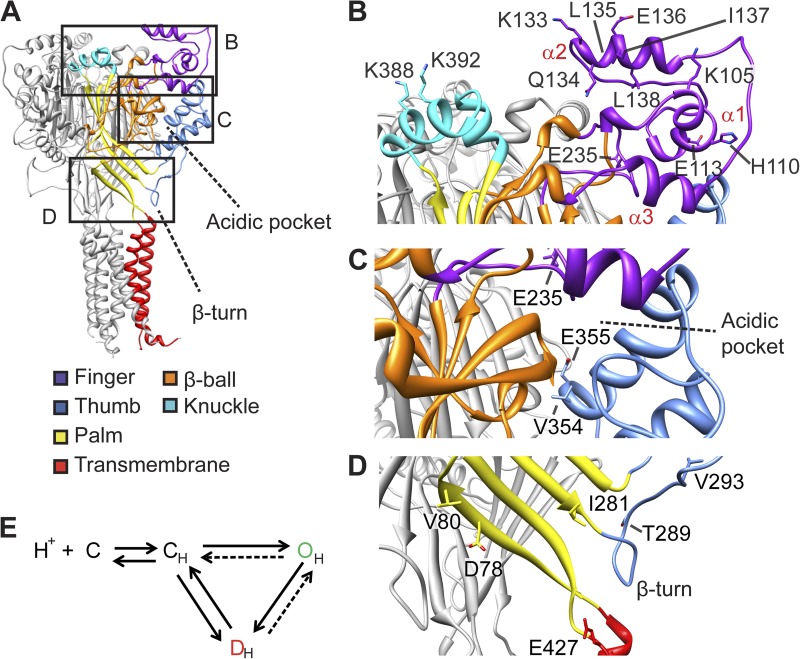
Figure 1.
Fluorophores placed at distinct sites of the ASIC protein report conformational changes. (A) Structural model of ASIC1a, based on the crystal structure of chicken ASIC1 (Jasti et al., 2007), with the domains in one subunit indicated by differential coloring. The frames indicate part of the structure shown in close-up view in B–D. (B–D) Detailed views indicating the localization of the residues that were mutated to Cys for labeling by fluorophores. (B) Finger and knuckle domains, (C) Acidic pocket, (D) palm and palm-thumb loop with β turn. (E) ASIC gating scheme. Protonation (CH) leads to a transient opening (OH) followed by channel desensitization (DH) or, upon mild acidification, to desensitization from the closed state (SSD).

