Figure 6.
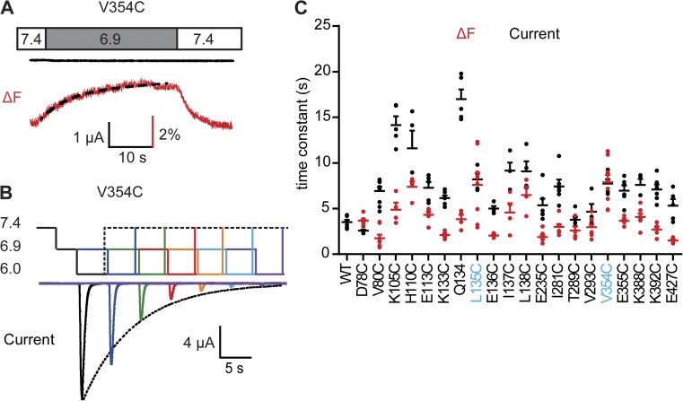
Fluorophores in the thumb and finger domain report movements related to SSD. (A) Representative experiment of an oocyte expressing the V354C mutant. The extracellular pH is changed as indicated from the conditioning pH 7.4 to the stimulation pH 6.9 that was not sufficiently acidic to activate the channels but generated a substantial ΔF. The onset of the ΔF was fitted to a single exponential (black dashed line). (B) Protocol applied to determine the SSD time course of ASIC1a. pH values corresponding to the different levels are indicated on the left. Channels were repeatedly activated by stimulation pH 6. Between stimulations, channels recovered during 40 s at pH 7.4. The pH 6.0 stimulation was directly preceded by incubation at conditioning pH 6.9 for a duration that increased in each round. The resulting current amplitude decrease, as a function of the incubation time at pH 6.9, was fitted to a single exponential (dotted line). (C) Time constants of the ΔF onset at stimulation pH 6.9 (red), and of the current SSD time course as determined in (B) at conditioning pH 6.9 (black), are shown for the different mutants; n = 4–10. For D78C and V80C, pH 7.0 was used instead of pH 6.9. Mutants whose ΔF is associated with SSD (i.e., their ΔF and current time constants are not statistically different [ANOVA and Tukey post-hoc test; P > 0.05], and their ratio is within the limit of 1.4-fold) are labeled in blue. Error bars represent SEM. All experiments were performed with fluorophore-labeled channels.

