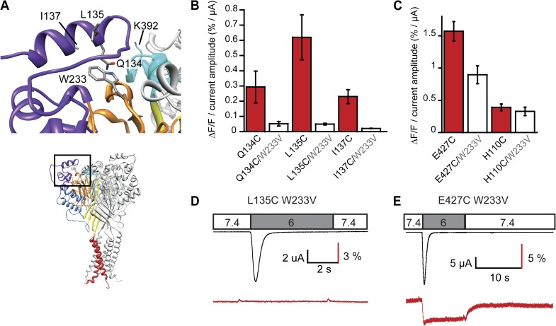
Figure 8.
The finger moves away from the β ball upon channel activation. (A) View of the finger residues Q134, L135, and I137, W233 of the β ball, and K392 of the knuckle of a neighboring subunit. (B) ΔF/F normalized to the acid-induced current amplitude is indicated for the three individual finger mutants and when each of them was combined with the W233V mutation (pH 6; n = 4–10). (C) ΔF/F normalized to the acid-induced current amplitude is indicated for two control mutations at a distance of >15 Å from W233V and when each of them was combined with the W233V mutation (pH 6; n = 7–8). (D and E) Representative current and ΔF trace of the double mutant L135C/W233V and the double mutant control E427C/W233V. Error bars represent SEM.

