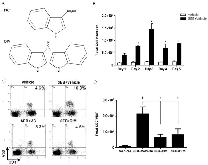
FIGURE 1. Treatment with I3C or DIM in vivo reduces percentage and number of SEB-specific Vβ8 T cells.
(A) Chemical structure of I3C and DIM. (B) C57BL/6 mice were given injections of 10ug of SEB in each hind footpad only once. Total cellularity from popliteal lymph nodes isolated from vehicle-treated versus SEB-treated mice was depicted. Mice were given ip injections of I3C or DIM (40mg/kg) for three consecutive days prior to SEB injection, which was followed by I3C and DIM treatment every other day. During the peak period of cell expansion on day 3, percentages (C) and total cell numbers (D) of CD3+Vβ8+ from popiteal lymph nodes were determined in each experimental group (n=5) using flow cytometry and antibodies for the respective markers. Statistical significance (p-value <0.05) was determined using GraphPad Prism analysis software with one-way ANOVA and Tukey’s multiple comparsion test (+ indicates significance compared to Vehicle group, and * indicates significance compared to SEB+Vehicle).

