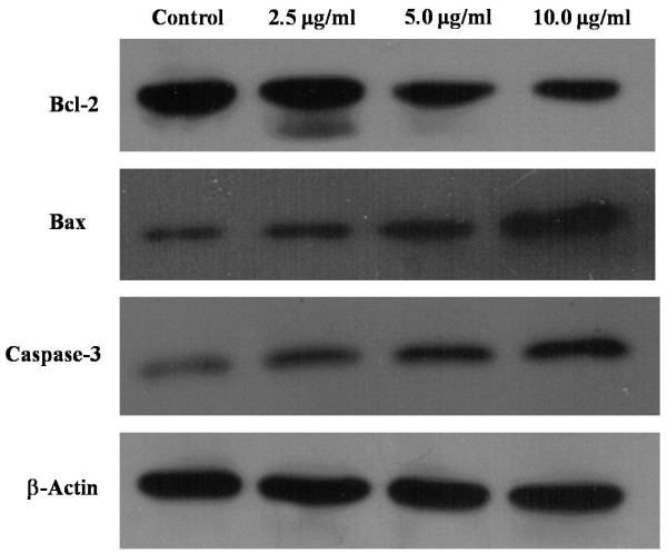
Figure 7.
Effect of ardipusilloside I on the expression of apoptotic proteins. Mc3 cells were exposed to various concentrations (2.5 μg/ml, 5 μg/ml, and 10 μg/ml) of ardipusilloside I for 48 h and the levels of Bcl-2, Bax, and caspase-3 were measured by Western blot analysis. Increased expression of Bax and caspase-3 and a decrease of Bcl-2 were observed. β-Actin was used as an internal loading control.

