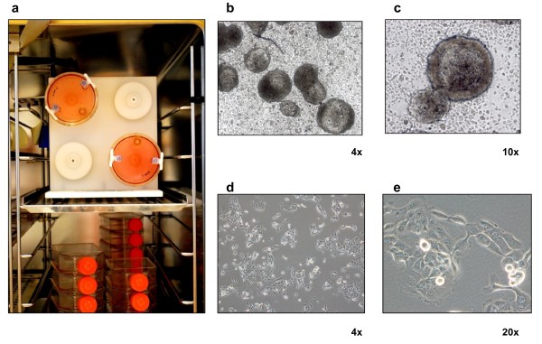
Figure 1.

Three-dimensional cell cultures. (a) MCTS growing in 50-mL vessels installed into the RCCS. Representative image of CAEP grown as (b, c) MCTS of about 1.3 -1.5 mm ∅ sampled from about 100 spherical colonies obtained from a single vessel or grown as (d, e) monolayer cultures with normal bright-field or phase contrast respectively, obtained by Olympus inverted microscope with attached Nikon high speed DS-Vi1 color digital camera. Spheroid number and size were determined through image analysis using NIS-Elements D software package.
