Figure 3.
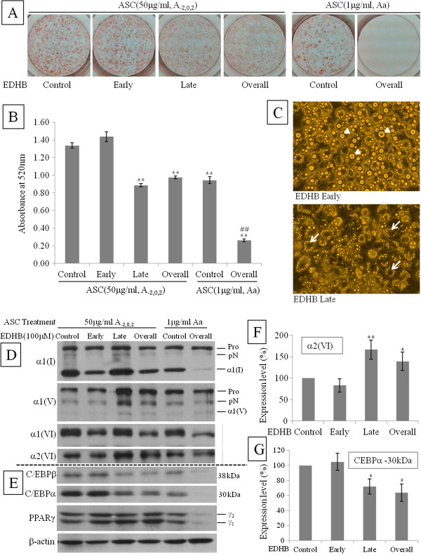
Inhibition of ASC in late phase removed lipid droplets from adipocytes. Cells were treated with high concentration (50 μg/ml) of ASC on early phase (A-2,0,2) or low concentration (1 μg/ml) of ASC on overall phase (Aa). EDHB (100 uM) was applied to ASC-treated cells on early, late, or overall phase. EDHB non-treated cells were regarded as control. Inhibitory effect was determined by ORO staining on Day 8 after chemical induction. After pictures (A) were taken, ORO stain was extracted and absorbance (B) was measured at 520 nm. Data are expressed as means ± SEM (bars). **p < 0.01 vs control of high ASC, ##p < 0.01 vs control of low ASC. (C) Microscopic determination of inhibitory effect. Arrow heads indicated large lipid droplets while arrows indicated the empty spots of large lipid droplet. (D) Immunoblot determination of inhibitory effect on collagens. Pro, procollagen; pN, N-procollgen; α1(I), mature α1 chain of type I collagen; α1(V), mature α1 chain of type V collagen. (E) Immunoblot determination of inhibitory effect on transcription factors. Expression level of a2(VI) (F) and CEBPa (30 kDa) (G) in high concentration of ASC (50 μg/ml, A-2,0,2) was semi-quantified. After adjusting control as 100%, band densities are expressed as means ± SEM (bars). *p < 0.05 and **p < 0.01 vs control.

