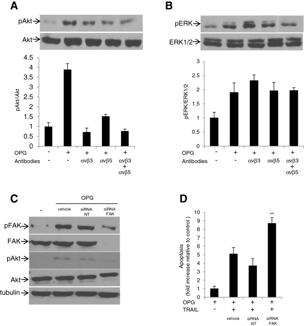
Figure 4.
Integrin/FAK mediates OPG-induced Akt activation. CaOV3 cells were incubated with αvβ3 and αvβ5 integrin blocking antibodies (5 μg/ml) for 1 h. Cells were washed, incubated with OPG (25 ng/ml) for 90 min and subsequently lysed for immunoblot with (A) anti-Akt and anti-phospho-Akt antibodies or (B) anti-ERK1/2 or anti-phospho-ERK1/2 antibodies. Densitometric quantification of phosphorylated Akt from three separate experiments normalized to total Akt was done. (C) CaOV3 cells were treated with either lipid vehicle alone, non-targeted siRNA (NT siRNA) or FAK siRNA for 24 h. OPG (25 mg/ml) was then added for 90 min and cells were subsequently lysed and immunoblotted for total FAK, phosphorylated FAK, total Akt and phosphorylated Akt. (D) CaOV3 cells were preincubated for 24 h with either lipid vehicle alone, non-targeted siRNA (NT siRNA) or FAK siRNA. OPG (25 ng/ml) was then added for 90 min. Cells were washed and incubated with TRAIL (50 ng/ml) for 24 h, and apoptosis was assessed. Apoptosis is expressed as fold increase relative to control (untreated) cells with the mean of triplicates from three independent experiments ± SD. * P < 0.01, ** P < 0.001 compared to NT siRNA treated cells.

