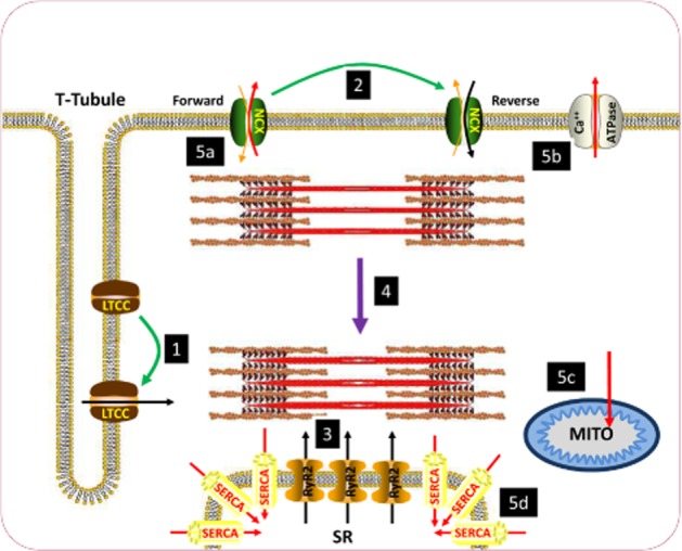
Figure 1.

Schematic diagram of excitation-contraction coupling in the ventricular cardiomyocyte. (1) Sarcolemmal depolarization causes L-type Ca2+ channels (LTCC) to open, resulting in influx of Ca2+ during systole. Ca2+ enters initially through the LTCC. (2) During the early stages of the action potential, the Na+–Ca2+ exchanger (NCX) also transitions from functioning in forward mode (Na+ in Ca2+ out) to reverse mode (Ca2+ in Na+ out) to further enhance the rise in [Ca2+]i. (3) The initial rise in [Ca2+]i induces a larger release of Ca2+ from the sarcoplasmic reticulum (SR) via ryanodine receptors (RyR2), resulting in a large rise in [Ca2+]i. (4) The large rise in Ca2+ causes contraction of the myofilaments. (5) [Ca2+] is subsequently reduced to bring about diastole through movement out of the cytosol (a) via NCX in forward mode and (b) the sarcolemmal Ca2+-ATPase. (c) Ca2+ is also buffered by mitochondria. (d) The majority of Ca2+ movement in diastole is into the SR via SERCA2a. Green arrows denote transitions brought about by sarcolemmal depolarization. Black arrows denote routes of influx of Ca2+ to the cytosol during systole. Purple arrow denotes contraction of myofilaments. Red arrows denote routes of efflux of Ca2+ during diastole.
