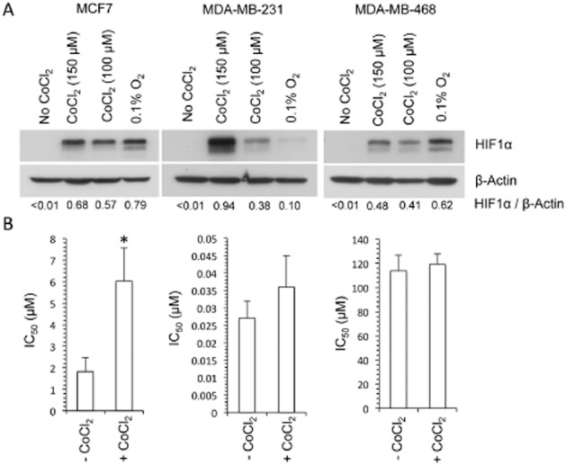
Figure 6.

The role of HIF1 in the activity of dasatinib. Western blot analysis of the effect of hypoxia (24 h exposure to 0.1% oxygen) and CoCl2 (6 h exposure to 150 μM and 24 h exposure to 100 μM CoCl2 under aerobic conditions) on the expression of HIF1α is presented in panel (A). The values presented below each lane represent the ratio of HIF1α to β-actin expression as determined by densitometry. The response of cells to dasatinib in the absence and presence of CoCl2 is presented in panel (B). Cells were pretreated with 150 μM CoCl2 for 6 h before a 1 h exposure to dasatinib and 150 μM CoCl2. Each value presented represents the mean ± SD for three independent experiments. Statistical significance (P < 0.01) between the control (no CoCl2) and the CoCl2-treated groups is indicated by *.
