
Figure 4.
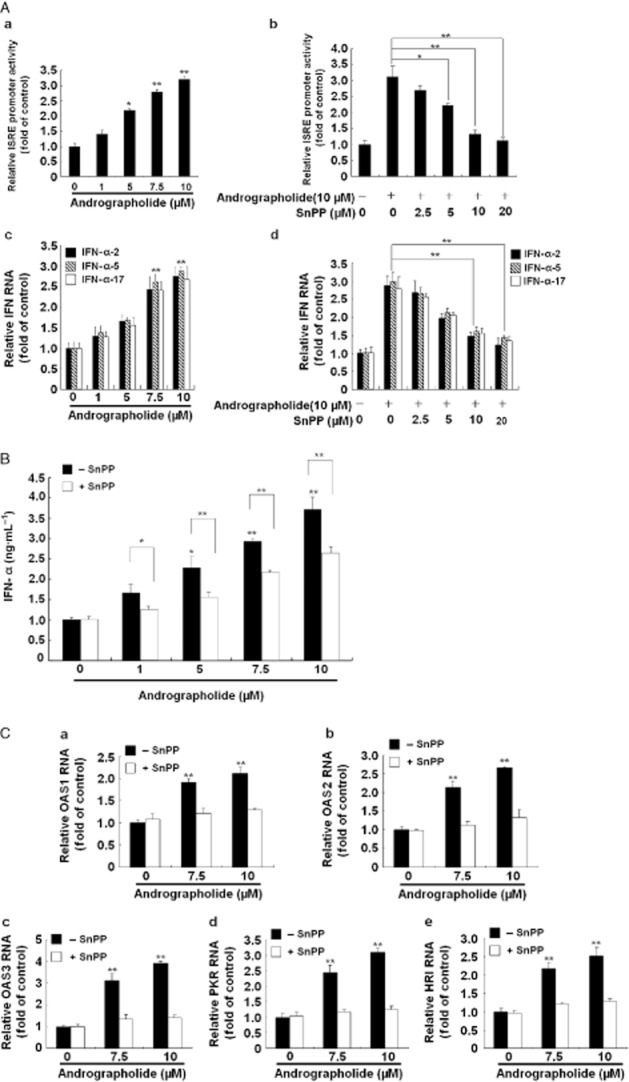
Andrographolide induces antiviral IFN responses via HO-1 induction. (A, panels a–d) Induction of ISRE activity (a) and gene expression of IFN-α-2, IFN-α-5 and IFN-α-17 (c) by andrographolide and attenuation of this effect by the HO-1-specific inhibitor SnPP (b and d). (B) Induction of IFN-α protein level by andrographolide and attenuation of this stimulant effect by SnPP. (C, panels a–e) Induction of IFN-mediated gene expression by andrographolide and suppression of this effect by SnPP. Ava5 cells or pISRE-Luc-transfected Ava5 cells were incubated with andrographolide alone at the indicated concentrations (0–10 μM) or coincubated with increasing concentrations of SnPP (0–20 μM) and 10 μM andrographolide for 3 days. The ISRE promoter activity was assayed for luciferase activity and was presented as fold activation relative to andrographolide/SnPP-untreated cells (defined as 1). The protein level of extracellular IFN-α was measured by elisa. The mRNA levels of IFN-α-2, IFN-α-5, IFN-α-17, OAS1, OAS2, OAS3, PKR or haeme-regulated inhibitor were measured by qRT-PCR and normalized against the cellular gapdh mRNA level and presented as fold change relative to the andrographolide/SnPP-untreated control (defined as 1). The experiments were performed in triplicate, and error bars indicate mean ± SD. *P < 0.05, **P < 0.01.