
Figure 5.
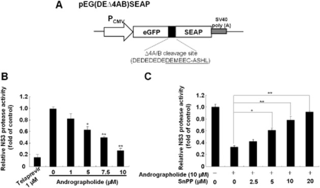
Andrographolide inhibits HCV NS3/4A protease activity via HO-1 induction. (A) Schematic diagram of the NS3/4A response reporter vector including the HCV cleavage site flanked by egfp and seap genes, named as pEG(DEΔ4AB)SEAP. (B) Andrographolide inhibited NS3/4A protease activity in a concentration-dependent manner. (C) Restoration of andrographolide-reduced NS3/4A protease activity by the HO-1-specific inhibitor SnPP. The pEG(DEΔ4AB)SEAP vector and the NS3/4A expression vector pNS3/4A were transiently co-transfected into Huh-7 cells. Each transfection mixture contained 0.1 μg pFLuc reporter plasmid as a control of transfection efficiency. The transfected cells were then treated with andrographolide alone at different concentrations (0–10 μM) or co-treated with increasing concentrations of SnPP (0–20 μM) and 10 μM andrographolide for 3 days. Telaprevir treatment served as the positive control. The NS3/4A protease activity was determined by SEAP activity following normalization against luciferase activity and presented as fold change relative to the andrographolide/SnPP-untreated control (defined as 1). The experiments were performed in triplicate, and error bars indicate mean ± SD. *P < 0.05, **P < 0.01.