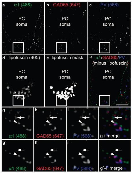
Figure 1.
Four channel imaging experimental design. Sections containing human dorsolateral prefrontal cortex (DLPFC) area 9 were imaged for (a) GABAA α1 subunit (488 channel), (b) glutamic acid decarboxylase 65 (GAD65; 647 channel), (c) parvalbumin (PV; 568 channel) and (d) lipofuscin autofluorescence, which is visible in all channels (405 channel). (e) Mask of the lipofuscin signal. (f) Merged image of remaining immunolabel used for analysis after lipofuscin subtraction. (g–i) 3X zoom of the boxed region showing fluorescent signal in the 488, 647 and 568 channels, respectively. (g'–i') 3X zoom of the boxed region showing fluorescent signal in the 488, 647 and 568 channels, respectively, after lipofuscin autofluorescence subtraction. Filled arrowhead indicates a triple-immunolabeled parvalbumin basket cell (PVBC) input included in final analysis. Filled arrow indicates a false-positive PVBC input (g–i merge) eliminated after lipofuscin autofluorescence subtraction (g'–i' merge). Scale bar: 5 μm. PC, pyramidal cell.

