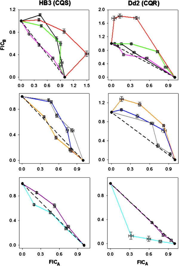
Figure 2.
IC50-based isobologram curves against HB3 (CQS, left column) and Dd2 (CQR, right column) for combinations with CQ (top), AQ (middle), and PQ/TQ (bottom) as “drug A” (x-axes). CQ-PQ (black), CQ-TQ (red), CQ-MB (green), CQ-AQ (pink), AQ-PQ (dark blue), AQ-TQ (orange), AQ-MB (gray), PQ-MB (purple), and TQ-MB (light blue or cyan). FICA and FICB correspond to the fractional inhibitory concentrations (see Methods) of the first and second drugs in each pair listed above, respectively. Error bars represent the standard error of the mean (S.E.M.) for duplicate experiments, each performed in triplicate (6 determinations total).

