
Figure 6.
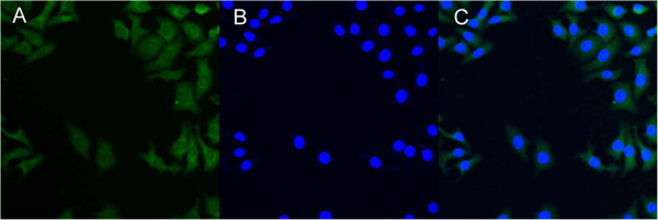
CLSM images of MCF-7 cells after 4 h of incubation with the coumarin 6-loaded CA-PLA-TPGS nanoparticles. The coumarin 6-loaded nanoparticles were green, and the cells were stained by DAPI (blue). The cellular uptake was visualized by overlaying images obtained using the EGFP filter and DAPI filter: (A) EGFP channel, green; (B) DAPI channel, blue; and (C) combined EGFP channel and DAPI channel.