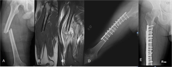
Figure 5.

A 37-year-old man with a history of type 2 diabetes mellitus. (A) Plain film of the femur showing a pathological fracture with lateral cortex thinning and laminated periosteal reaction. (B, C) T1- and T2-weighted magnetic resonance imaging (MRI) showing severe soft tissue swelling and edematous change. (D) Plain film taken two months after open reduction and internal fixation (ORIF) and debridement surgery showing recurrent osteomyelitis with loss of reduction and laminated periosteal reaction of the medial cortex. (E) Plain film taken one year after revision debridement and ORIF surgery showing bony union without signs of osteomyelitis recurrence.
