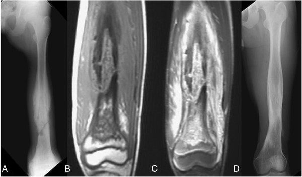
Figure 6.

A 14-year-old boy who experienced moderate progressive thigh pain, especially at night, for four months. (A) A plain film of the femur showing a laminated periosteal reaction with pathological fracture at the distal femur. (B, C) Magnetic resonance imaging (MRI) showing tumor-like lesion at the marrow surrounded with extensive soft tissue edema. (D) A plain film of the femur taken two years after casting and debridement surgery showing bony union.
