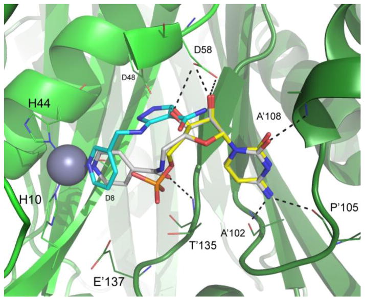
Figure 2.
Crystal structure of compound 3 (carbon atoms white) bound to BpIspF (PDB 3KE1) depicting the active site comprised of chains A (light green ribbon) and B (dark green ribbon). Overlaid are binding poses of fragments FOL8395 (carbon atoms cyan) and cytidine monophosphate (carbon atoms yellow) from PDB entries 3JVH and 3F0G, respectively10 Ligand oxygen atoms are colored red, nitrogen blue, and phosphorus orange. Active site residues critical for ligand binding with high conservation across pathogenic organisms are indicated. Residues 64–72 from chain A were unmodeled due to poor electron density in the diffraction data. Residues 60–63 were removed from image for clarity. Image created using PyMol 14.

