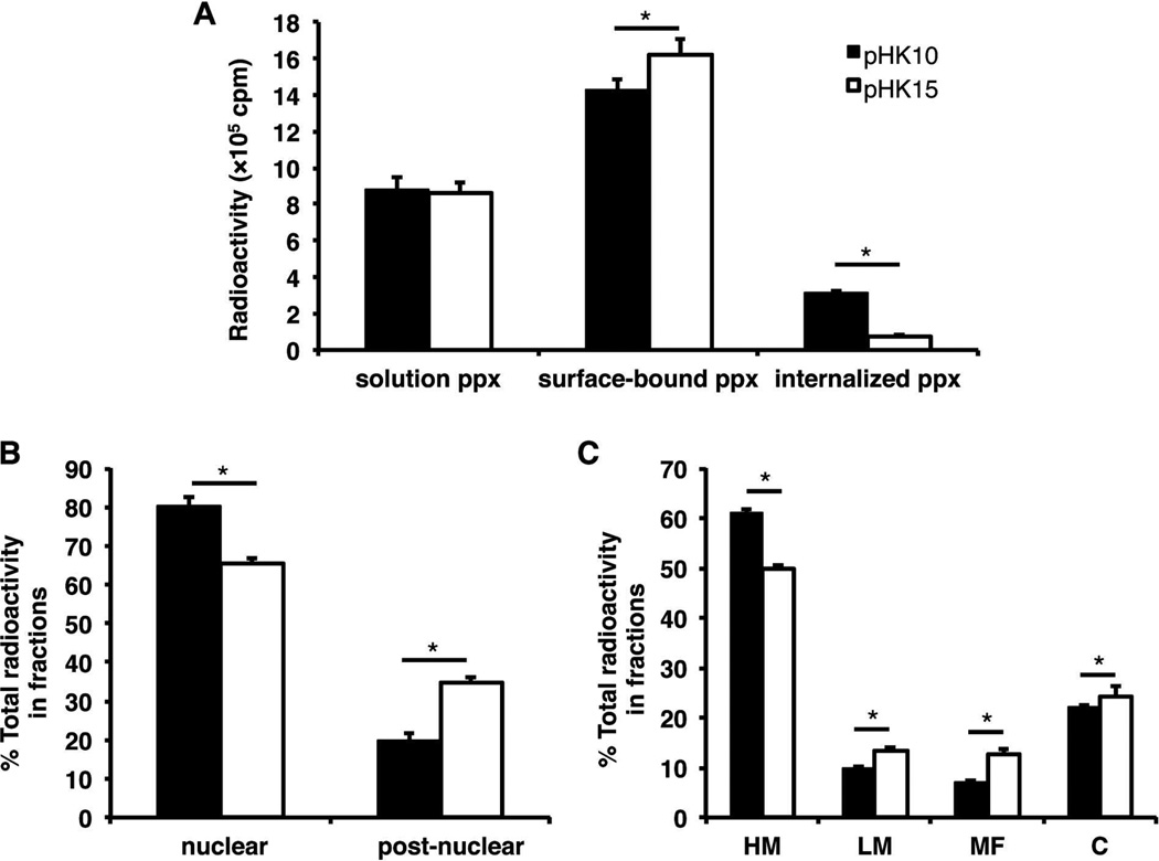
Figure 7.
Subcellular distribution of [3H]DNA/polymer complexes in HeLa cells. HeLa cells (5 × 10⁶) were treated with pHK10 (black bars) or pHK15 (white bars) polyplexes (containing 100 µg DNA) for 4 h prior to being washed with CellScrub to remove extracellularly-bound polyplexes and fractionated into nuclear, heavy mitochondrial (HM), light mitochondrial (LM), microsomal (MF), and cytosolic (C) fractions. (A) The amount of [3H]DNA found in solution (pulse), surface-bound (CellScrub), and internalized (trypsinized cells) fractions, (B) nuclear and post-nuclear fractions, and (C) post-nuclear fractions, composed of HM, LM, MF, C fractions. Data are presented as mean ± S.D., n = 3. (*) denotes p ≤ 0.05, as determined by a two-tailed, unpaired Student’s t-test with unequal variance.

