
Figure 2.
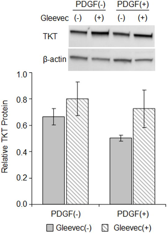
Antagonizing PDGF-receptor alters PDGF-induced loss of TKT. RCK cultured for 4 days in serum-free medium without [PDGF(-)] or with 100 ng/ml PDGF-BB [PDGF(+)] in conjunction without [(-), solid bars] or with [(+), striped bar] 10 μM ST1571 (Gleevec). Representative immunoblot of cytoplasmic lysates probed for TKT and beta-actin (top panel). Graph of immunoblot data (bottom panel). Relative TKT protein, a ratio of the TKT to the beta-actin band densities for each immunoblot (n=2), expressed as average ± high/low. TKT bands are ∼70 kD and beta-actin bands are ∼42 kD based on molecular size markers (not shown).