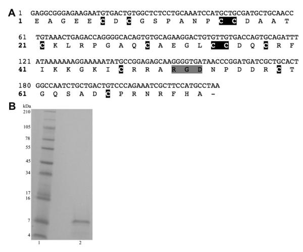
Fig. 1.
A) cDNA sequence and predicted amino acid sequence of r-viridistatin 2. The cDNA sequence is located on the upper line and the amino acid sequence on the lower line. The cysteine residues are shaded in black and the RGD motif is shaded in gray. B) SDS Gel electrophoresis. r-Viridistatin 2 was run reduced on a 10–20% Tricine gel. The gel was run with 1× Tricine SDS running buffer using an XCell SureLock Mini-cell at 125 V for 90 min. The gel was stained with Simply Blue Safe Stain for 1 h and destained with Milli-Q water. Lane 1) See Blue Plus 2 markers; Lane 2) r-viridistatin 2 (20 μg).

