
Fig. 1.
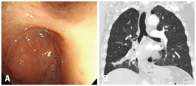
Bronchoscopic examination of a hamartoma shows a round shape, smooth and rough surface, pinkish color, and no visible vessels on the tumor surface. Spontaneous bleeding was absent (A). Chest computed tomography shows a 25-mm calcified ovoid nodule at the distal bronchus intermedius and atelectasis of the right middle lobe (B).