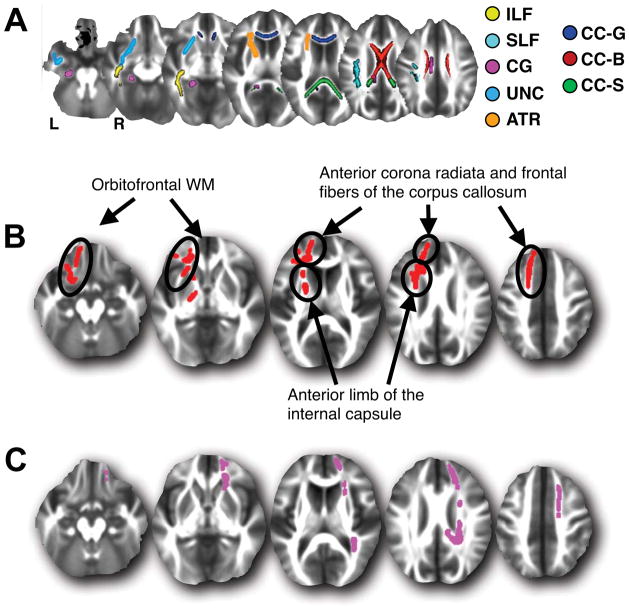
Figure 1.
(A) Skeletal WM regions of interest (ROI). Areas are shown in the left hemisphere only except for regions of the corpus callosum. Inferior and superior longitudinal fasciculi (ILF and SLF), cingulate gyrus (CG), uncinate fasciculus (UNC), anterior thalamic radiation (ATR), corpus callosum genu (CC-G), body (CC-B) and splenium (CC-S). (B) Areas where lower reasoning scores are associated with higher skeletal AD after accounting for the effects of age and gender. (C) Areas where lower flexibility scores are associated with higher skeletal AD after accounting for the effects of PS, age and gender. Depicted slices in (B) and (C) are the same and correspond to MNI z-values of -16mm, -2mm, 12mm, 26mm and 40mm.

