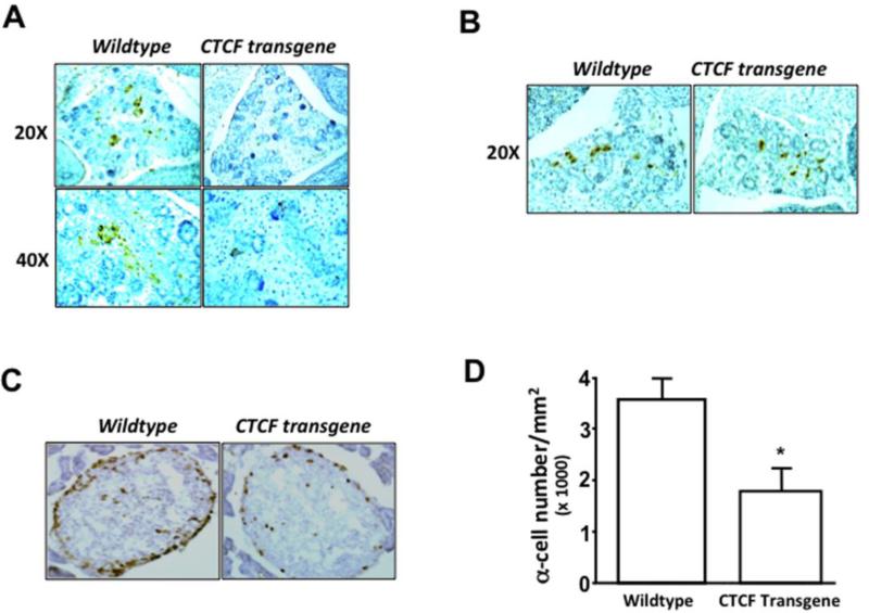
Figure 1. Effect of over-expressing CTCF on mouse islet α cell development.
(A) Immunostaining of pro-glucagon-containing cells in pancreatic islets. (B) Immunostaining of insulin-containing cells in pancreatic islets. Tissue sections were prepared from embryonic pancreases of sibling wild-type and CTCF transgenic mice at day 14 (E14). Pancreatic tissues were stained with anti-glucagon and anti-insulin antibodies. (C) Identification of pro-glucagon-positive α cells in pancreatic islets of adult mice by immunostaining with anti-glucagon antibodies. (D) Comparison of α cell populations in pancreatic islets of sibling wildtype and CTCF transgenic mice. Cell numbers were quantified by measuring pro-glucagon-positive cells per mm2. Pancreatic tissue sections were prepared from 4-5 months old wildtype and CTCF transgenic mice. Symbol “*” represents a significant difference (p<0.05, n=15).

