
FIG. 5.
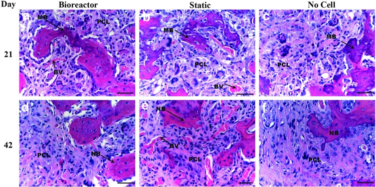
40×objective images of H&E stained defect implanted with PLGA/PCL scaffolds after 3 weeks (a–c) and 6 weeks (d–f ) of in vivo implantation. Before implantation scaffolds were cultured in vitro in the TPS bioreactor with an hMSC population (a, d), in static culture with an hMSC population (b, e), or in static culture with no cell population (c, f ). In images (a, b) note mineralization formation and blood vessel infiltration within PLGA/PCL scaffold. In image (c) note mineralized bone formation around the edge of the scaffold. Bone in growth continues to penetrate scaffold after 42 days (d–f ). Note minimal tissue response to material. Scale bar represents 50 μm. PCL, PLGA/PCL scaffold; NB, new bone; MB, mineralized bone; BV, blood vessel.