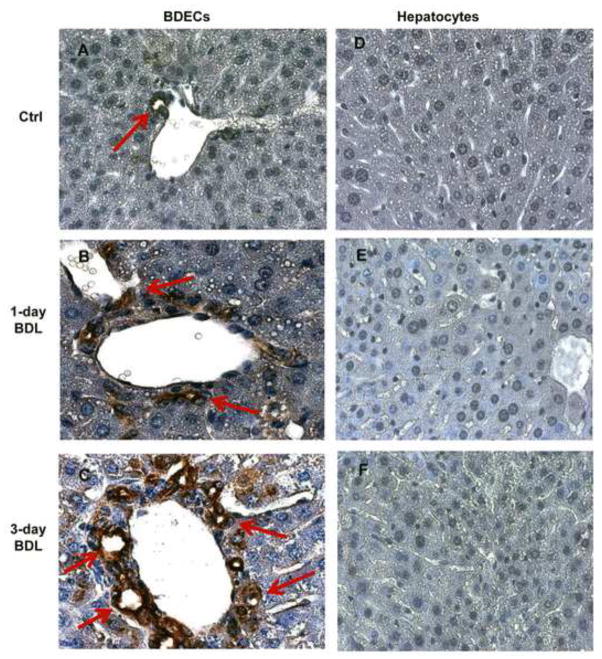
Figure 4.
OPN expression in bile duct epithelial cells (BDECs) and hepatocytes. WT animals were subjected to BDL for 1 or 3 days and liver section were stained by immunohistochemistry for OPN using an anti-OPN antibody. BDECs show increased expression of OPN after BDL (B, C). While there was extensive staining of BDECs (red arrows), no OPN expression was observed in hepatocytes (D, E, F). Magnification, x100.

