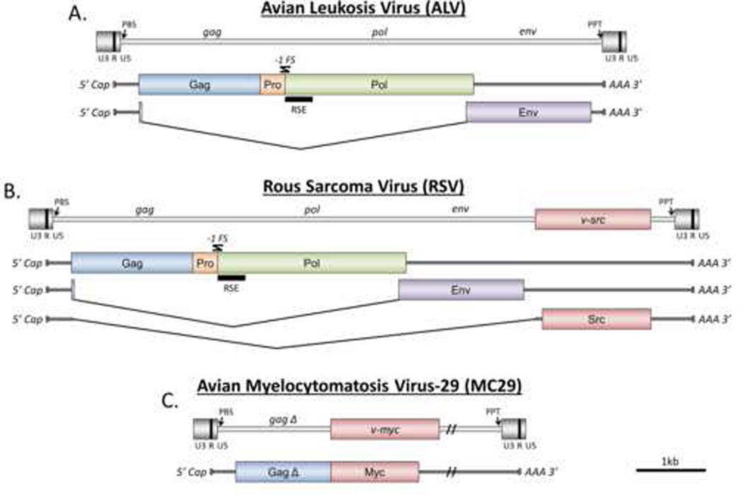
Figure 1.
The ASLV proviruses and the transcripts of ALV (A), RSV (B), and MC29 (C) are shown. The location of viral oncogenes within the proviral genome are indicated when they exist. Protein coding portions of the transcripts are represented by colored rectangles. Locations of primer binding sites (PBS), RNA stability elements (RSE), polypurine tracts (PPT), and −1 framshift sites (−1 FS) are indicated. Polyprotein cleavage sites are not shown. This figure was adapted from [62].

