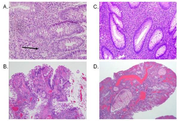
Figure 1.
Mild chronic colitis and juvenile polyp with adenomatous changes in Patient 1 with JP/HHT: A. Mixed inflammatory infiltrate in the lamina propria, focally invading the glandular epithelium (arrow). Mild architectural distortion, tortuosity of the glands was present. B. Juvenile polyp with focal adenomatous changes characterized by hyperchromasia and pseudostratification of the epithelial nuclei. Patient 2 with JP/HHT: C. Dense mixed inflammation including numerous eosinophils in the lamina propria. Mild architectural distortion with glandular trifurcation was seen. D. Juvenile polyp with ectatic vessels from colectomy specimen.

